Taxonomy: f. Hexamitidae
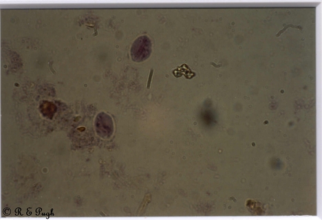
Animal: Giardia intestinalis 08 24.jpg
Sites: Gut
Comment:
Giardia intestinalis cysts - Haematoxylin stain. Giardia cysts are oval - ellipsoidal or round shape, 8 - 19 (usually 11-14) x 7-10 microns with thick wall; 4 nuclei with large central karyosome and no peripheral chromatin; remains of locomotor apparatus, deep staining median bodies lie across longitudinal fibers. Often there is shrinkage with cytoplasm pulling away from wall and also may have a halo effect around outside of wall.Although these cysts can be identified in wet smears, many infections are missed without the examination of a permanent stained smear. Also, because the trophozoites of Giardia are attached so firmly by means of their sucking discs in the mucosa; a series of even 5 or 6 samples may be examined without detecting the organisms. Incubation time for giardiasis is 12-20 days; the acute stage is brief but foul smelling, explosive diarrhoea plus lack of blood ,mucus or cellular exudate is consistent with giardiasis. A sub acute or chronic stage may continue with epigastric pain, abdominal distension, belching and weight loss and episodes of foul stools (Garcia & Bruckner, 1997).
First Picture |
Previous Picture |
Next Picture |
Last Picture

